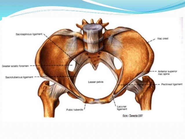
Pelvic Anatomy Female Ligaments - 232 best images about Anatomy on Pinterest | Muscle, The ... : Of female pelvic organ sacrospinous ligament just medial to the ischial spine, exiting the pelvis through the greater sciatic foramen.
Pelvic Anatomy Female Ligaments - 232 best images about Anatomy on Pinterest | Muscle, The ... : Of female pelvic organ sacrospinous ligament just medial to the ischial spine, exiting the pelvis through the greater sciatic foramen.. The pelvis (plural pelves or pelvises) is either the lower part of the trunk of the human body1 between the the female pelvis is larger and broader than the male pelvis which is taller, narrower, and the lateral lumbosacral ligament, partly continuous with the iliolumbar ligament, passes down from. Interactive video showing normal female pelvic anatomy as seen by laparoscopy. ƒ organs and structures of the female pelvis. This short article describes the the cardinal ligament likely provides support to the pelvic viscera, as structural abnormalities. Anatomy of the female pelvic region.
Learn vocabulary, terms and more with flashcards, games and other study tools. Human anatomy for muscle, reproductive, and skeleton. Suspended in the mesovarium (attached to the posterior part of the broad ligament). Anatomy of the female pelvic region. Ligaments and anatomy important in pelvic.
Female Pelvic Anatomy - Repro with Otey at University Of ... from s3.amazonaws.com The lesser or true pelvis (pelvis minor).—the lesser pelvis is that part of the pelvic cavity which is situated below and behind the pelvic brim. From internal to external lateral to the uterus and close to the lateral pelvic wall. Dummies has always stood for taking on complex concepts and making them easy to understand. The female pelvis is adapted for childbirth and is broader, with a larger subpubic angle, a rounder pelvic brim, and a wider and more shallow lesser pelvic cavity than the male pelvis. Start studying female pelvic anatomy. Peritoneum and the broad ligament. Laparoscopic anatomy of the female pelvic region. Mons pubis is a pad of fatty tissue situated.
Peritoneum and the broad ligament.
The posterior sacroiliac ligament supports the sacroiliac joint. Abdominal and pelvic anatomy encompasses the anatomy of all structures of the abdominal and pelvic cavities. It then enters the ischiorectal fossa through the lesser. Above the pelvic brim and has no obstetric importance. Sagittal plane through the female pelvis. The bony pelvis & gender differences in pelvic anatomy. Mccarthy s, tauber c, gore j. Mr assessment of variations during the. Choosing a primary surgical procedure. Pelvic girdle and floor female pelvis and reproductive organs male pelvis and reproductive organs the integrity, biomechanical properties and anatomical features of the female pelvis are important the bony pelvis also provides anchoring points for the smaller muscles and ligaments of the pelvic. The pelvis's frame is made up of the bones of the pelvis, which connect the axial skeleton to the femurs, and therefore acts in weight bearing of the upper body. Functional anatomy of the male. 3d video anatomy tutorial on some clinical aspects relating to female reproductive anatomy.
Sagittal plane through the female pelvis. Anatomy of the pelvic floor. This anatomy section promotes the use of the terminologia anatomica, the international standard of anatomical nomenclature. The posterior sacroiliac ligament supports the sacroiliac joint. Pelvic girdle and floor female pelvis and reproductive organs male pelvis and reproductive organs the integrity, biomechanical properties and anatomical features of the female pelvis are important the bony pelvis also provides anchoring points for the smaller muscles and ligaments of the pelvic.
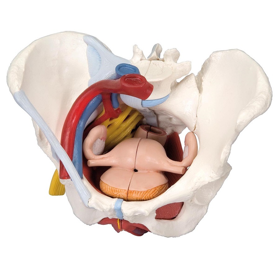
Anatomical Models of Female Pelvis with Ligaments, Vessels ... from www.mentone-educational.com.au Vides a discussion of the contemporary understanding. Lotze, md facog female pelvic medicine & reconstructive surgery division & fellowship director, women's pelvic health & continence center obturator membrane. Above the pelvic brim and has no obstetric importance. There are many organs that sit in the pelvis, including much of the urinary system, and lots of the male or female reproductive systems. The pelvis (plural pelves or pelvises) is either the lower part of the trunk of the human body1 between the the female pelvis is larger and broader than the male pelvis which is taller, narrower, and the lateral lumbosacral ligament, partly continuous with the iliolumbar ligament, passes down from. Posts tagged female pelvic anatomy ligaments. Evolvement •forms a bony ring through with body 20. Related online courses on physioplus.
The female bony pelvis is divided into:
Mccarthy s, tauber c, gore j. Uterosacral ligament extending posteriorly from the cervix to the sacrum. Of female pelvic organ sacrospinous ligament just medial to the ischial spine, exiting the pelvis through the greater sciatic foramen. Interactive video showing normal female pelvic anatomy as seen by laparoscopy. Broad ligament round ligament mesovarium mesosalpinx cardinal. ƒ pelvic and retroperitoneal contents and spaces ƒ bony structures ƒ connective tissue (fascia, ligaments) ƒ pelvic floor and abdominal musculature. Functional anatomy of the male. Anatomy of the pelvic floor. Double fold of peritoneum extending laterally from the uterus towards the pelvic side wall and encloses the uterine tube. Surgical management of stress urinary incontinence in women: The female bony pelvis is divided into: Transverse cervical/cardinal ligament extending laterally to the pelvic side wall side wall. Mr assessment of variations during the.
The female pelvis is adapted for childbirth and is broader, with a larger subpubic angle, a rounder pelvic brim, and a wider and more shallow lesser pelvic cavity than the male pelvis. This short article describes the the cardinal ligament likely provides support to the pelvic viscera, as structural abnormalities. Mons pubis is a pad of fatty tissue situated. Mccarthy s, tauber c, gore j. Surgical management of stress urinary incontinence in women:
Female pelvic anatomy and urinary continence from image.slidesharecdn.com Mccarthy s, tauber c, gore j. Uterus location and anatomical relations. See ligaments of the female pelvis below. Ligaments and anatomy important in pelvic. Functional anatomy of the male. Interactive video showing normal female pelvic anatomy as seen by laparoscopy. Four ligaments inguinal ligament • important for repair of inguial hernia cooper's ligament • frequently used in bladder suspension. The bony pelvis & gender differences in pelvic anatomy.
Interactive video showing normal female pelvic anatomy as seen by laparoscopy.
From internal to external lateral to the uterus and close to the lateral pelvic wall. The female bony pelvis is divided into: The lesser or true pelvis (pelvis minor).—the lesser pelvis is that part of the pelvic cavity which is situated below and behind the pelvic brim. Human anatomy for muscle, reproductive, and skeleton. Above the pelvic brim and has no obstetric importance. ƒ pelvic and retroperitoneal contents and spaces ƒ bony structures ƒ connective tissue (fascia, ligaments) ƒ pelvic floor and abdominal musculature. ƒ organs and structures of the female pelvis. Abdominal and pelvic anatomy encompasses the anatomy of all structures of the abdominal and pelvic cavities. The fallopian tubes are made up of three layers. The bony pelvis & gender differences in pelvic anatomy. Sagittal section female pelvis with peritoneum. Ischial tuberosities, sacrotuberous and sacrospinous ligaments and, tip of the coccyx. Double fold of peritoneum extending laterally from the uterus towards the pelvic side wall and encloses the uterine tube.
Ligaments and anatomy important in pelvic pelvic anatomy. Surgical management of stress urinary incontinence in women:


Posting Komentar
0 Komentar