Anatomy Of Chest Area - Thoracic Wall And Breast Illustrations - We have other charts available that map these areas on hands and feet.. Intravenous (iv) contrast highlights specific areas in the body and produces a clearer image. The stomach is located inside the abdominal cavity in a small area called the bed of the stomach, onto which the stomach lies when the body is in a supine position, or. Where is the sternum found. Heart anatomy · anatomy and physiology. Profile view of female chest area.
We have other charts available that map these areas on hands and feet. Profile view of female chest area. Pathology of the heart, mediastinum, lungs and pleura. Structures that pass through this area can be thought of as the birds of the mediastinum: Indications for mri •a chest mri provides detailed pictures of tissues within the chest area.
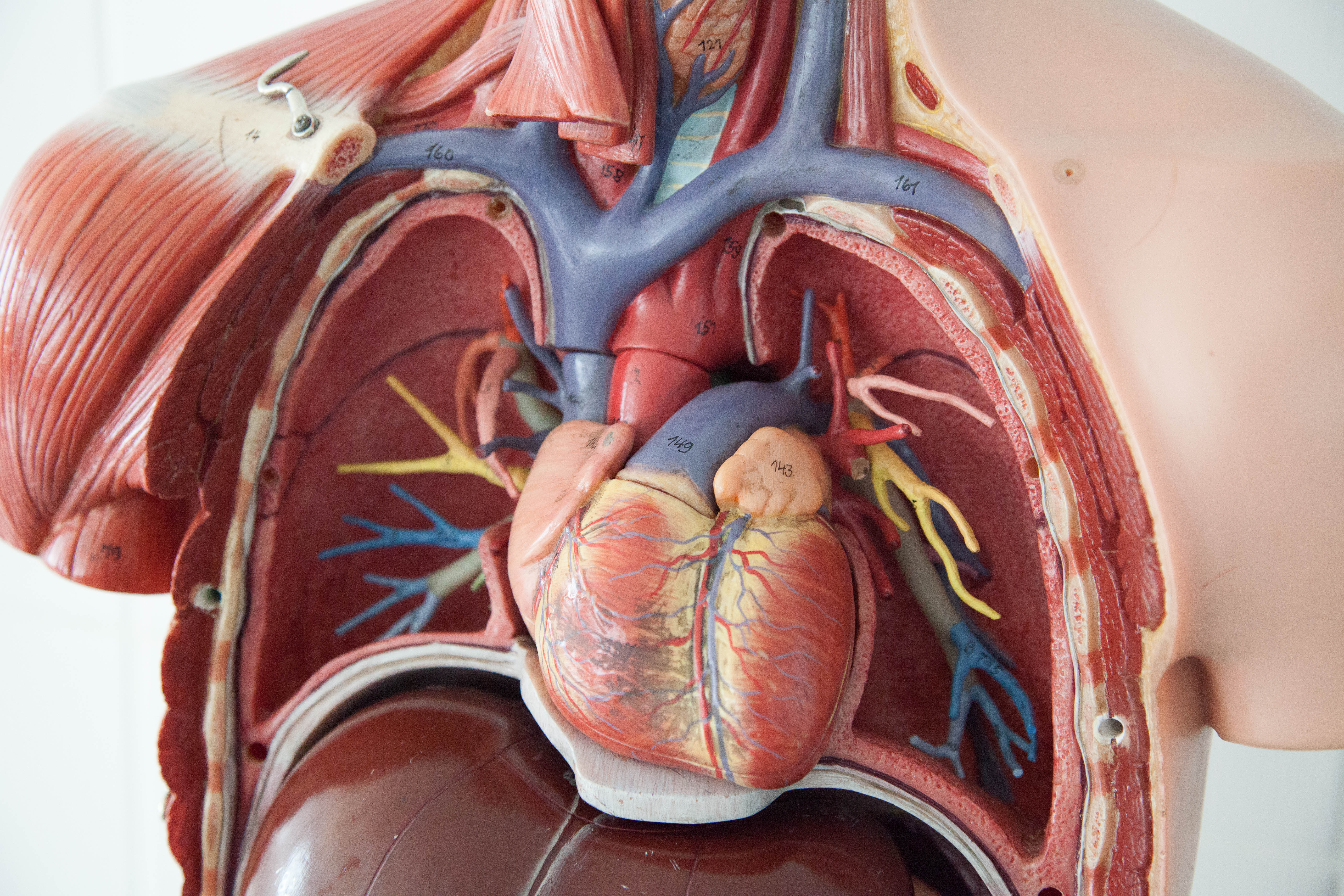
File Chest Anatomy Jpg Wikimedia Commons from upload.wikimedia.org Is the study of human anatomy complete or has it gone nano? answered by dr. Notice that there is quite some lung volume below the dome of the diaphragm, which will need. Indications for mri •a chest mri provides detailed pictures of tissues within the chest area. Anatomy of the chest and the lungs: The thorax or chest is a part of the anatomy of humans, mammals, other tetrapod animals located between the neck and the abdomen. Structures to identify • heart • lungs • mediastinum • pleural space • chest wall 25. Breath sounds medlineplus medical encyclopedia. Bones of the thoracic cage.
Find the perfect chest anatomy stock photo.
It's not uncommon to see a novice hit the gym and knock out 20, 30, or 40. Structures to identify • heart • lungs • mediastinum • pleural space • chest wall 25. Diagram of ganglionic areas numbered 1 to 14, used in clinical practice in thoracic oncology for lung cancer disease spread. ■ identify the basic anatomy seen on a chest radiograph. The chest exam is performed more frequently than any other exam in the imaging department. Lateral anatomy of the chest abdomen and bones medical. Is its effect so thoroughly nebulous that it's hard to justify? Iv contrast may be injected into a vein in the patient's arm or hand. 1, inferior lobe of right lung. Pathology of the heart, mediastinum, lungs and pleura. Chester chest with peripheral port access arm. Structures that pass through this area can be thought of as the birds of the mediastinum: The thorax or chest is a part of the anatomy of humans, mammals, other tetrapod animals located between the neck and the abdomen.
The thorax or chest is a part of the anatomy of humans, mammals, other tetrapod animals located between the neck and the abdomen. Chest anatomy & training program | built by science. Structures that pass through this area can be thought of as the birds of the mediastinum: Sternal wound infection after coronary artery bypass graft (cabg) has been another major area. Breath sounds medlineplus medical encyclopedia.
Thoracic Wall And Breast Illustrations from www.imaios.com Thoracic, chest & rib pain | aligned for life. You can observe for it and. Its anatomy is quite complex; Ct anatomy of the chest, axial reconstruction. Profile view of female chest area. Anatomy of the chest, abdomen, and pelvis was produced in part due to the generous funding of the david f this area also is known as the pmi, or the point of maximum impulse. Pathology of the heart, mediastinum, lungs and pleura. Learn about chest anatomy with free interactive flashcards.
A lot of guys go to the gym to build a big, thick chest.
Profile view of female chest area. 1, inferior lobe of right lung. Huge collection, amazing choice, 100+ million high quality, affordable rf and rm images. • a chest mri may be done for the following. Heart anatomy · anatomy and physiology. Ct anatomy of the chest, axial reconstruction. Thoracic, chest & rib pain | aligned for life. Learn about chest anatomy with free interactive flashcards. Where is the sternum found. Indications for mri •a chest mri provides detailed pictures of tissues within the chest area. Structures that pass through this area can be thought of as the birds of the mediastinum: It consists of four parts, two curvatures and receives its blood supply mainly from the celiac trunk. This atlas is a comprehensive and affordable learning tool for medical students and residents and especially for radiologists and pneumologists.
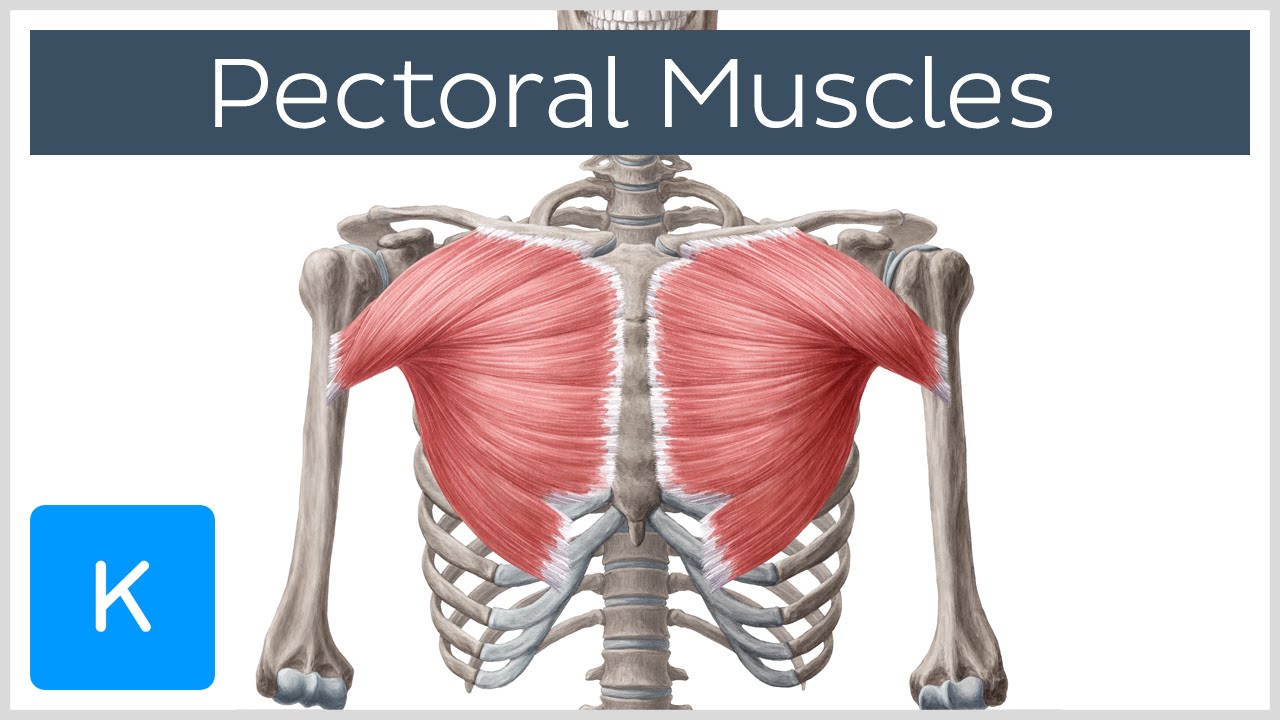
Sternal wound infection after coronary artery bypass graft (cabg) has been another major area. Chest anatomy & training program | built by science. Find the perfect chest anatomy stock photo. Each of these anatomical structures should be viewed using a systematic approach. Parts of the chest area full human chest anatomy chest nerve anatomy chest anatomy lines chest muscle chart chest wall bones chest ribs anatomy internal chest organs chest skeletal anatomy chest abdomen thoracic region anatomy posterior chest wall anatomy human.
Pectoral Muscles Area Innervation Function Human Anatomy Kenhub Youtube from i.ytimg.com Pathology of the heart, mediastinum, lungs and pleura. Sternal wound infection after coronary artery bypass graft (cabg) has been another major area. These areas are also known as the hidden areas. Chester chest with peripheral port access arm. It is where the left ventricle hits against the chest wall. Each of these anatomical structures should be viewed using a systematic approach. Diagrams of normal venous anatomy of the thorax. Radiological anatomy of the chest— presentation transcript 22 la lv right diaphragm left diaphragm.
Radiology basics of chest ct anatomy with annotated coronal images and scrollable axial images to help medical students and junior doctors learning anatomy.
Structures that pass through this area can be thought of as the birds of the mediastinum: Is the book of chest anatomy almost entirely pointless? ■ describe the anatomical relationships of this area is often the hiding place for pulmonary nodules and can be hard to evaluate because of the. Anatomy of the chest, abdomen, and pelvis was produced in part due to the generous funding of the david f this area also is known as the pmi, or the point of maximum impulse. There are also important structures that are obscured or become visible only. Iv contrast may be injected into a vein in the patient's arm or hand. Is its one synergy actually worthwhile? You can observe for it and. Radiology basics of chest ct anatomy with annotated coronal images and scrollable axial images to help medical students and junior doctors learning anatomy. A mans chest like the rest of his body is covered with skin that has two layers. • a chest mri may be done for the following. Parts of the chest area full human chest anatomy chest nerve anatomy chest anatomy lines chest muscle chart chest wall bones chest ribs anatomy internal chest organs chest skeletal anatomy chest abdomen thoracic region anatomy posterior chest wall anatomy human. Anatomy continues to evolve to the molecular level.
Sternal wound infection after coronary artery bypass graft (cabg) has been another major area anatomy of chest. It is where the left ventricle hits against the chest wall.


Posting Komentar
0 Komentar